


Instrumentation Laboratory
- Home
- Faculty of Engineering & Technology
- Department of Physics and Nanotechnology
- Instrumentation Laboratory
Specifications
Current Equipments
| Name of the Equipment and Make | Broad Specifications | Equipment |
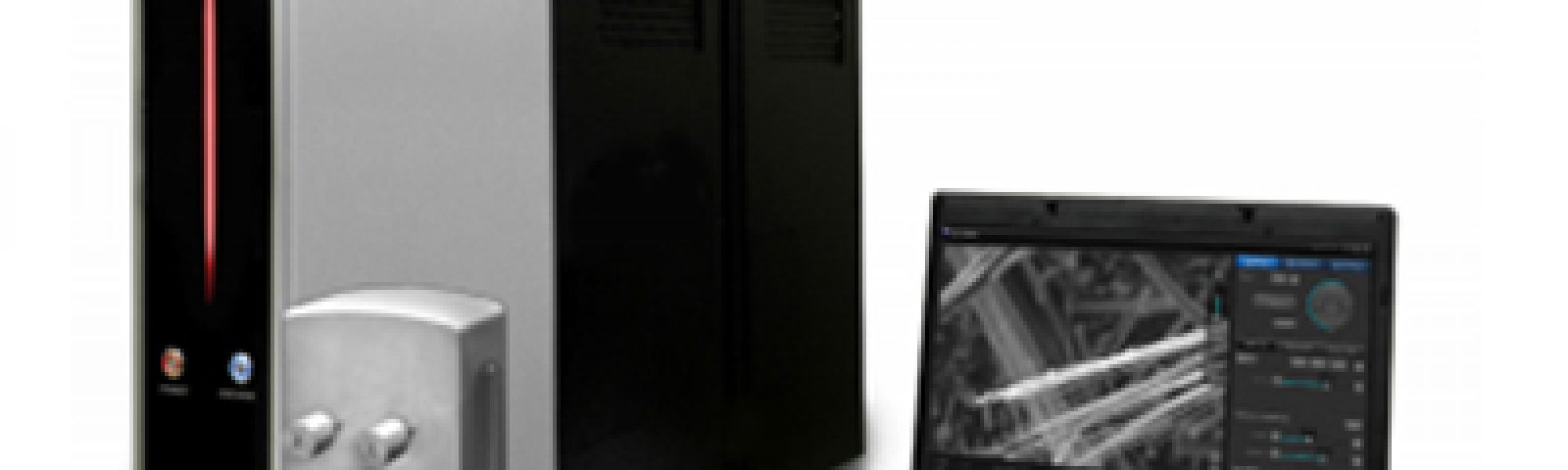
| DESKTOP Mini-SEM with EDS
(SNE- 3200M, SEC)
|
Max. 30,000x Magnification
BSE Detector (Solid State Type – 4 Channel), Resolution (SE): ~ 15.0nm@30kV, Resolution (BSE): ~ 20.0nm @30Kv, 5kV to 30kV Variable Accelerating Voltage, Multi-Vacuum Mode – Standard / Charge Up Reduction, Image Observation Ready within 3 min., 3-axis Strokes – X, Y, R (Option – X, Y, T),Options – EDX System, Cooling Stage |
 |
| CLASSIC LINEPULVERISETTE 5
FRITSCH |
Planetary Mill classic line PULVERISETTE 5 instrument without
grinding bowls and balls, incl. Safe-Lock clamping system with 2 grinding bowl fasteners for 100-120/200-240 V/1~, 50-60 Hz, 250Watt Voltage, indicated by customer is set Hardmetal tungsten carbide, with steel casing. Grinding bowls 80 ml volume. Adapter for grinding bowl of 80 ml volume. Hardmetal tungsten carbide 10 mm balls. Replacement seal ring PTFE 90/75 mm dia. for all other bowls of 80 ml volume(spare).
|
 |
| KEITHLEY System Source meter – Dual Channel
|
200V: LOW CURRENT Dual Channel,6-1/2 digits display, 60 Watts,
Current Max / Min: 10A pulse / 0.1fA, Voltage Max / Min: 200V / 100nV,GPIB, LAN (LXI), RS-232, USB, Digital I/O |

|
| OLYMPUS MODEL BX51 Upright Metallurgical Microscope
|
Microscope frame for reflected & transmitted light microscopy,
Trinocular tube,Objective 50X,Incident bright field/dark field illumination tube with aperture 1 No. stop, field stop and filter slots,Olympus High resolution 2.8MP Microscope digital camera system with USB 3.0 interface
|
 |
| Scanning Probe Microscope
Park Systems |
XY Scanner-Scan range: 50μm x 50μm;
10μm x 10μm Motorized Z Stage, Direct On-Axis Optical Microscope, High Resolution Digital CCD Camera with Digital Zoom, Manual Focus Stage for On-Axis Optics, Motorized Z Stage, Software-XEP for data acquisition and optical view, XEI for image processing, analysis, and presentation. Standard Imaging (AFM Modes): True Non-Contact AFM, Basic Contact AFM, Lateral Force Microscopy (LFM), Phase Imaging. Intermittent (tapping) AFM-Scanning Tunneling Microscopy, Conductive AFM, Nanolithography, Magnetic Force Microscopy, Nano indentation, Force Modulation Microscopy. |
 |
|
Electrochemical Work Station, SP-300 from BioLogic
|
Potentiostat/Galvanostatmeasurements, Corrosion:
Linear/cyclic polarization, corrosion, pitting, LPR, ZRA(noise), RpvsTime,Impedance: potentio EIS, Galvano EIS, Staircase Potentio EIS, GalvanoEIS.Current ranges: 1 A to 1 μA. Pulsed techniques.
|
 |
| TD-NMR, BRUKER
|
Frequency range: (Direct-digital generation)2-65 MHz with
1 Hz stepping and better than 1 ppm resolution. Digital phase generation: 0°, 90°, 180°, and 270°, with phase resolution better than 0.2.Digital quadrature receiver with variable gain: 40- 119 dB.Magnetic system: 20 MHz, 470 mTeslaminispec magnet system with 25 mm air gap and fully integrated and automated temperature. Magnet Temperature Control: 35 °C to 45 °C.Probe Assembly: Variable High & Low Temperature proton probe at 20 MHz suitable for ratio measurements for sample tubes with 10 mm diameter. Glass Sample Tubes 10 mm diameter, 180 mm length, flat bottom. Proton probe at 20 MHz suitable for absolute measurements (Total Fat, Spin Finish) for sample Bath: Thermostat/Cryostat Bath -10 to +100 °C
|
 |
|
X-ray Fluorescence Spectroscopy Panalytical
|
Model: Minipal 4 Benchtop XRF.
Elemental range: Al…Y, Pd…U. Size: 300x550x450 mm3.Fine focus X-ray tube with MO Target. Multilayer monochromator 17.5 Kev
|
 |
| X’ pert powder XRD system
Panalytical
|
Radiation-Co Kα,Goniometer Type: Vertical, Range of 2θ: 0-150°.Detector Type: High speed solid-state X’Celerator Optics: Divergence and antiscattering slits. Sample holders:Flat Silicon ZBH Cut parallel to Si (510), Back-fill holder
|
 |
| UV-Visible Spectroscopy
UV 3000+ from LABINDIA |
Double beam optics (Czerny-Turner mounting),
Wavelength range: 190 to 1100nm. Spectral bandwidth: variable 0.5,1,2,5 nm. Wavelength accuracy: ±0.3nm. Photometric accuracy: ±0.002 Abs at 0.5 Abs. Photometric repeatability: ±0.001 Abs at o.5 Abs. Automatic 8 cell changer. Tungsten and deuterium lamp. Microprocessor based UV-VIS Spectrophotometer with high resolution. LCD display and soft keypad for operation on 220V/50Hz
|
 |
| FTIR Spectroscopy
|
Spectral range: 500-7500 cm-1.
Spectral resolution: Better than 2cm-1.Wave number accuracy: Better than 0.01 cm-1.Detector: DTGS. Source: SiCglobar, user replaceable. Rock Solid interferometer: Gold mirrors (permanently aligned). Holder: 2×3” standard sample mount & also for pellets. Accessory required for ATR setup.
|
|
|
AAS7000 Atomic Absorption Spectrophotometer LABINDIA
|
Fully automatic double beam PC
controlled high performance with window 2000/XP based wizard software for operation on 220V/50 Hz. Monochromator: Czereny-Turner type. Wavelength range 190-900 nm. Sealed, corrosive resistant and vibration free optical system. Reciprocal linear dispersion less than 1.6 nm/mm.Detector: Photomultiplier tube.6 Lamp motorized turret. Burner system: Air Acetylene type. Hollow Cathode Lamps: 6. Sensitivity: An absorbance of 0.9 or better, for 5ppm Cu solution.
|
 |

No data was found
No data was found
No data was found
No data was found
No data was found
No data was found
No data was found
No data was found